Our promise to you:
Guaranteed product quality, expert customer support.
 24x7 CUSTOMER SERVICE
 CONTACT US TO ORDER
LAG-3 Gene Editing 
Lymphocyte activation gene 3 (LAG-3), a member of the immunoglobulin superfamily, is expressed in tumor infiltrating lymphocytes, B cells, NK cells, dendritic cells, and has the function of binding to MHC molecules of class II present in tumor cells. LAG-3, one of the main checkpoint receptors that coordinate the regulation of regulatory T cells and anergic T cells, through its interplay with the class II molecule of MHC results in the downregulation of the proliferation of antigen-specific CD4+ lymphocytes. LAG-3 expression has been observed in various types of cancer, such as melanoma, colorectal cancer, breast cancer, Hodgkin's lymphoma, ovarian cancer, multiple myeloma, hepatocellular carcinoma, chronic lymphocytic leukemia, and gastric cancer.
LAG-3 Signaling
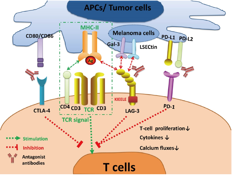
Following TCR engagement, Lag-3 associates with CD3 in the TCR complex and crosslinking of Lag-3 together with CD3 negatively regulates signal transduction resulting in reduced T cell proliferation and cytokine production. However, due to the lack of a definable motif in the cytoplasmic tail, the molecular aspects of the inhibitory effects of Lag-3 are still largely unknown. The cytoplasmic tail of Lag-3 does not contain any inhibitory motifs shared with other inhibitory receptors. Therefore, the exact signaling pathway utilized by Lag-3 is still unclear. However, the Lag-3 cytoplasmic tail contains three regions that are conserved between human and mouse, which may be involved in signal transduction. The first region contains a serine-phosphorylation site, the second region contains a single lysine residue in a unique "KIEELE" motif, and the third region contains glutamic acid-proline (EP) repeats. Among these three regions, the KIEELE motif has been proved to be necessary for signal transduction and Lag-3 inhibition.
 Figure 1. LAG-3 signaling and the interaction with other immune checkpoints. (Long L, et al., 2018)
LAG3 may also mediate bidirectional signaling into the interacting APCs. MHC class II binding to LAG3-expressing Tregs has been shown to inhibit DC activation, thereby suppressing their maturation. CD86 upregulation was inhibited along with the decrease of IL-12 secretion mediated by an ITAM inhibitory signaling pathway involving FcγRγ and ERK-mediated recruitment of SHP-1. This is indeed a reverse signaling mechanism as a LAG3 mutant without the cytoplasmic tail was enough to suppress DCs function. LAG3-expressing Tregs may utilize this mechanism to enhance tolerance by indirectly inhibiting DC function. LAG3 has a similar reverse signaling mechanism in the interaction between DC and melanoma cells. When exposed to LAG3-transfected cells, MHC class II-expressing, but not MHC class II-negative, melanoma cells were resistant to Fas-mediated apoptosis by activating MAPK/ERK and PI3K/Akt survival pathways.
Targeting LAG-3 in Cancer Immunotherapy
Consistently, the levels of LAG-3 expression and infiltration of LAG-3+ cells in tumors have been reported to be related to the tumor progression, poor prognosis, and unfavorable clinical outcomes in various types of human tumors, such as colorectal cancer, follicular lymphoma, breast cancer, renal cell carcinoma, head and neck squamous cell carcinoma (HNSCC), non-small cell lung cancer, and diffuse large B cell lymphoma. These results strongly suggest that LAG-3 contributes to immune escape mechanisms in tumors similar to PD-1. Therefore, LAG-3 has been considered as a promising therapeutic target for cancer immunotherapy, which is also supported by studies using animal models.
The anti-LAG-3 antibody can not only promote the activity of effector T cells, but also inhibit Treg-induced suppressive function in the TME. In addition, in light of the interaction between LAG-3 and other immune checkpoints, targeting LAG-3 along with other checkpoints especially PD-1 holds considerable promise in cancer immunotherapy. So far, at least 13 agents that target LAG-3 have been developed. Anti-LAG-3 blocking Abs (relatlimab (BMS-986016), TSR-033, MK-4280, LAG525, Sym022, INCAGN2385-101, and BI754111) and antagonistic bispecific Abs (FS118 (anti-LAG-3/PD-L1), MGD013 (anti-PD-1/LAG-3), and XmAb22841 (anti-CTLA-4/LAG-3)) are under clinical trials for various cancers either as monotherapy or in combination primarily with anti-PD-1 or anti-PD-L1 blocking Abs.
LAG-3 Gene Editing Services
CRISPR/Cas9 PlatformCB, one of the leading biotechnological companies specializing in gene editing, is dedicated to offering comprehensive CRISPR/Cas9 gene-editing services to a wide range of genomics researchers. Based on our platform, we can help you effectively LAG-3 gene deleted, inserted or point mutated in cells or animals by CRISPR/Cas9 technology.
- LAG-3 Gene Knockout: We offer LAG-3 gene knockout cell line and knockout animal model generation service with high quality. Typically, we develop CRISPR-mediated gene editing cell lines including HEK239T, Hela, HepG2, U87, but we can use other cell lines according to your requirements. Our one-stop KO animal model generation service covers from sgRNA design and construction, pronuclear microinjection to Founders genotyping and breeding.
- LAG-3 Gene Knockin: CRISPR/Cas9 PlatformCB provides the one-stop LAG-3 knock-in cell line and knockout animal model generation services, including point mutation and gene insertion. Our expert staff has succeeded in dozens of LAG-3 knock-in cell line generation projects, including stem cells, tumor cells and even difficult-to-handle cells. We also have extensive experience in incorporating CRISPR/Cas9 technology into animal models, which have been fully recognized by our clients.
If you have any questions, please feel free to contact us.
Related Products at CRISPR/Cas9 PlatformCB
| CATALOG NO. | PRODUCT NAME | PRODUCT TYPE | INQUIRY |
| CDKM-0012 | B6J-Lag3em1Cflox | Knockout Mouse | Inquiry |
| CLKO-1304 | LAG3 KO Cell Lysate-HeLa | Knockout Cell Lysate | Inquiry |
| CSC-RT1454 | Human LAG3 Knockout Cell Line-HeLa | Pre-Made Knockout Cell Line | Inquiry |
References
- Andrews L P, et al. LAG 3 (CD 223) as a cancer immunotherapy target. Immunological reviews, 2017, 276(1): 80-96.
- Perez-Santos M, et al. LAG-3 antagonists by cancer treatment: a patent review. Expert opinion on therapeutic patents, 2019, 29(8): 643-651.
- Joller N, Kuchroo V K. Tim-3, Lag-3, and TIGIT. Emerging Concepts Targeting Immune Checkpoints in Cancer and Autoimmunity, 2017: 127-156.
- Long L, et al. The promising immune checkpoint LAG-3: from tumor microenvironment to cancer immunotherapy. Genes & cancer, 2018, 9(5-6): 176.
- Shan C, et al. Progress of immune checkpoint LAG‑3 in immunotherapy. Oncology Letters, 2020, 20(5): 1-1.
- Maruhashi T, et al. LAG-3: from molecular functions to clinical applications. Journal for Immunotherapy of Cancer, 2020, 8(2).