Our promise to you:
Guaranteed product quality, expert customer support.
 24x7 CUSTOMER SERVICE
 CONTACT US TO ORDER
CRISPR Rabbits for Ocular Disease Research 
The development of novel therapies for ocular diseases requires the development of animal models of ocular diseases with similar etiology, pathology, and suitability for future trials of new treatments. Compared to mice, rabbits are closer to humans in terms of phylogeny, anatomical features, physiology, and pathophysiological responses. Rabbits possess relatively large eyes and share many anatomical features with humans. These similarities include eye size, internal eye structure, eye optical system, eye biomechanics, and eye biochemical characteristics. In addition, rabbits have a relatively long lifespan, making the rabbit model ideal for studying age-related aspects of retinal degenerative diseases as well as long-term evaluation of treatments and side effects.
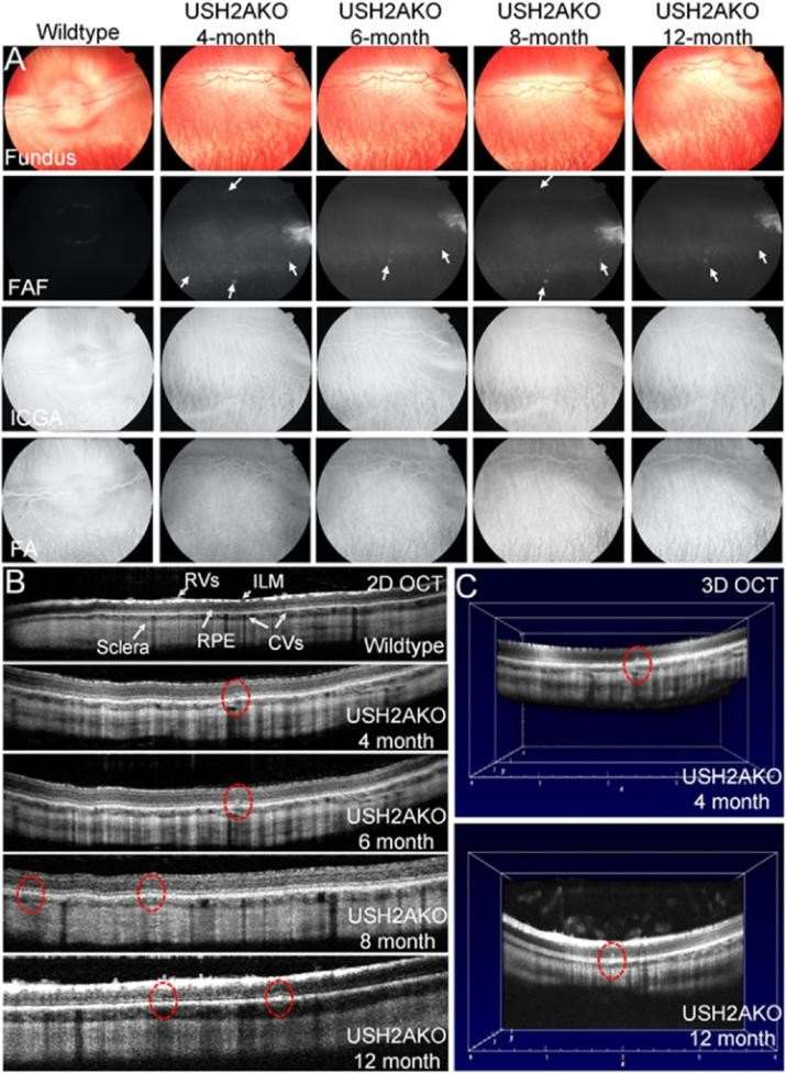
 Fig. 1 Retinal imaging in USH2A KO rabbit. (Nguyen VP, et al., 2023)
Solution
Creative Biogene has extended gene targeting to rabbits via CRISPR/Cas9 technology to develop gene targeting rabbit models that replicate inherited human eye diseases. Our existing knockout rabbit models include GJA8 knockout rabbits (congenital cataract model), αA-crystallin knockout rabbits (congenital cataract model), and USH knock-in and knock-out rabbits (Usher syndrome model). Among them, the congenital cataract model can summarize the phenotypes of congenital cataract, microphthalmia, early lens atrophy, and failure of lens fiber differentiation. the Usher syndrome model can summarize the phenotypes of progressive retinal photoreceptor degeneration and hearing loss.
Related Analytical Services
- Rabbit eye examination and imaging: We use slit lamp biomicroscopy to perform a thorough examination of the rabbit's eyelids, conjunctiva, cornea, anterior chamber, iris, and lens prior to imaging. We used fundus photography, fundus autofluorescence (FAF), fluorescein angiography (FA), and indocyanine green angiography (ICGA) to evaluate the vascular network of the retina and choroid.
- Electroretinographic analysis: We analyzed the visual function of rabbits under dark and bright vision conditions.
- We examined the neuroretinal conditions of rabbits by scanning electron microscopy, including the nuclear layer, plexiform layer, photoreceptors, retinal vasculature (RV), choroidal vasculature (CV), RPE, inner limiting membrane, and sclera.
- We performed the histopathological analysis of eye sections from rabbits to detect whether the photoreceptors of retinitis pigmentosa degenerated.
Related Services
Related Products
CRISPR/Cas9 PlatformCB is dedicated to providing you with gene-edited rabbit models for preclinical studies of common and rare ocular diseases, including DES, glaucoma, AMD, light-induced retinopathy, cataract and uveitis, diabetic retinopathy, retinal detachment and proliferative vitreoretinopathy, ocular allergy, retinoblastoma and Retinitis pigmentosa and other eye diseases. Our gene editing technology has greatly enhanced the value of rabbits in biomedicine, and our model promises to greatly facilitate basic and translational research in ocular diseases. If you are interested in our services, please feel free to contact us.
Reference:
- Nguyen VP, et al. USH2A Gene Mutations in Rabbits Lead to Progressive Retinal Degeneration and Hearing Loss. Transl Vis Sci Technol. 2023, 12(2):26.