Our promise to you:
Guaranteed product quality, expert customer support.
 24x7 CUSTOMER SERVICE
 CONTACT US TO ORDER
CD33 Gene Editing 
CD33, a 67-kDa glycosylated transmembrane protein, is a member of the sialic acid binding, immunoglobulin-like lectin family. In addition to committed myelomonocytic and erythroid progenitor cells, it is expressed on most myeloid and monocytic leukemia cells. It is not seen on the earliest pluripotent stem cells, lymphoid cells, mature granulocytes, or nonhematopoietic cells.
CD33 Function
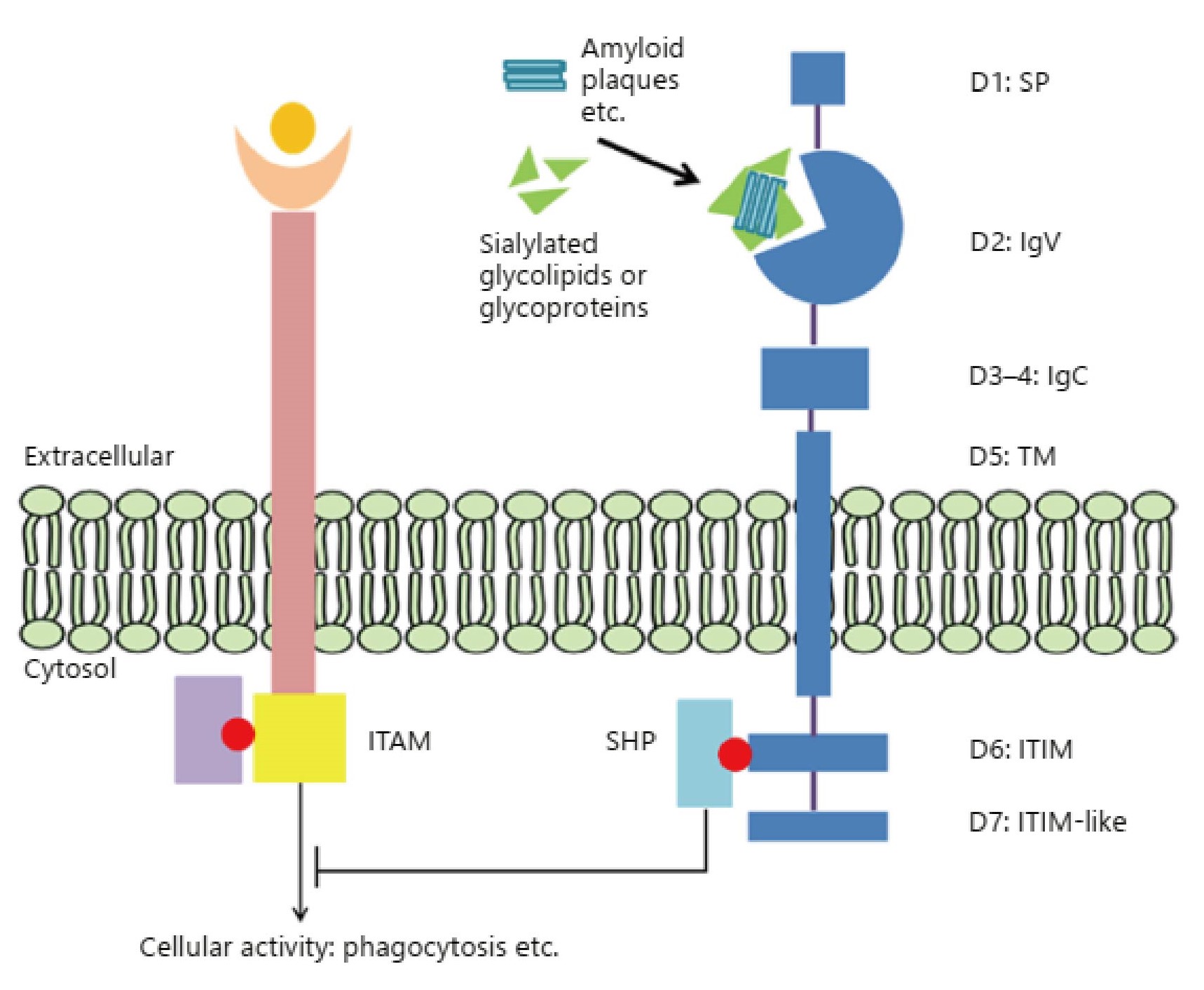
Due to the exclusive expression of CD33 on immune cells, an important role of CD33 is its ability to modulate immune cell functions. In immune cells, cellular processes (such as cytokine release, phagocytosis, apoptosis, etc.) are coregulated by immunoreceptor tyrosine-based activation motif (ITAM)-containing activating receptors and ITIM-containing inhibitory receptors. When binding to its ligands, the tyrosine in the ITIM of inhibitory receptors is phosphorylated and serves as a docking site for Src homology (SH) 2 domain-containing proteins like SHP phosphatases. This results in dephosphorylation of cellular proteins, which consequently downregulate activating signaling pathways involving phosphorylation, such as those induced by ITAMs. CD33 is an inhibitory receptor that can be constitutively activated by sialic acid-containing glycoproteins and glycolipids. The activated CD33 receptor recruits inhibitory proteins such as SHP phosphatases through its ITIM domains, leading to an inhibition of cellular function such as phagocytosis. CD33 is also involved in several other processes, such as inhibition of cytokine release by monocytes, adhesion processes in immune or malignant cells, immune cell growth and survival through the inhibition of proliferation, and induction of apoptosis.
 Figure 1. Schematic depiction of CD33 exon structure and its inhibitory mechanism. (Zhao L. 2019)
CD33 as a Target for Immunotherapy
CD33 is expressed on the cell surface of more than 80% of leukemia isolates from patients with acute myeloid leukemia (AML), and the average antigen density is very high. Surface levels can show considerable inter-patient variability. The differential expression of CD33 on the surface of malignant AML blasts, but not on the surface of normal hematopoietic stem cells (HSCs), makes CD33 an ideal target for immunotherapy. The most famous CD33-targeted immunotherapy that is clinically approved is the anti-CD33 antibody gemtuzumab ozogamicin (GO; Mylotarg), which is conjugated to the cytotoxic drug calicheamicin. GO was approved in 2000 for the treatment of patients over 60 years with CD33+ AML in first relapse who did not meet the requirements of standard chemotherapy, but was voluntarily withdrawn from the market when a confirmatory post-approval clinical trial was prematurely terminated because of lack of clinical benefit and increased adverse events. However, GO was reapproved in 2017 when further studies showed that it would be beneficial to add GO to standard chemotherapy if fractionated doses of the drug were used. Some immunotherapies targeting CD33, including antibody-drug conjugates, CAR T cells and bispecific antibodies, are currently under clinical evaluation or preclinical development.
CD33 as a Potential Therapeutic Target for AD
The amyloid-beta peptide (Aβ) cascade hypothesis posits that the accumulation of Aβ is the fundamental initiator of Alzheimer's disease (AD). Besides, it is generally believed that impaired Aβ clearance is a major pathogenic event for LOAD. Microglial cells play important role in Aβ clearance in the brain, and the recent study from Griciuc et al. showed that CD33 contributed to the pathogenesis of AD by impairing microglia-mediated clearance of Aβ. Moreover, deletion of CD33 both in vitro and in vivo resulted in the increased Aβ clearance and reduced Aβ levels. Remarkably, CD33 deletion in vivo was viable, because CD33 knockout mice were fertile and exhibited no anatomical defect, which means that targeting CD33 may be a safety option in AD treatment.
CD33 Gene Editing Service
CRISPR/Cas9 PlatformCB, one of the leading biotechnological companies specializing in gene editing, is dedicated to offering comprehensive CRISPR/Cas9 gene-editing services to a wide range of genomics researchers. Based on our platform, we can help you effectively CD33 gene deleted, inserted or point mutated in cells or animals by CRISPR/Cas9 technology.
- CD33 Gene Knockout: We offer CD33 gene knockout cell line and knockout animal model generation service with high quality. Typically, we develop CRISPR-mediated gene editing cell lines including HEK239T, Hela, HepG2, U87, but we can use other cell lines according to your requirements. Our one-stop KO animal model generation service covers from sgRNA design and construction, pronuclear microinjection to Founders genotyping and breeding.
- CD33 Gene Knockin: CRISPR/Cas9 PlatformCB provides the one-stop CD33 knock-in cell line and knockout animal model generation services, including point mutation and gene insertion. Our expert staff has succeeded in dozens of CD33 knock-in cell line generation projects, including stem cells, tumor cells and even difficult-to-handle cells. We also have extensive experience in incorporating CRISPR/Cas9 technology into animal models, which have been fully recognized by our clients.
If you have any questions, please feel free to contact us.
Related Products at CRISPR/Cas9 PlatformCB
References
- Clark M C, Stein A. CD33 directed bispecific antibodies in acute myeloid leukemia. Best Practice & Research Clinical Haematology, 2020: 101224.
- Zhao L. CD33 in Alzheimer's disease–biology, pathogenesis, and therapeutics: a mini-review. Gerontology, 2019, 65(4): 323-331.
- Jiang T, et al. CD33 in Alzheimer's disease. Molecular neurobiology, 2014, 49(1): 529-535.
- Estus S, et al. Evaluation of CD33 as a genetic risk factor for Alzheimer's disease. Acta neuropathologica, 2019: 1-13.
- Laszlo G S, et al. The past and future of CD33 as therapeutic target in acute myeloid leukemia. Blood reviews, 2014, 28(4): 143-153.
- Jurcic J G. What happened to anti-CD33 therapy for acute myeloid leukemia?. Current hematologic malignancy reports, 2012, 7(1): 65-73.